Is Medical Imaging The Same As Diagnostic Imaging?

Our Medical Imaging and Diagnostic Imaging are The Same?
You might have heard health practitioners throwing around words like “medical imaging” and “diagnostic imaging.” This might leave you confused and wondering whether these two terms are the same is not. To clear the air, let’s address this question. Both words mean precisely the same thing and will be discussed in detail in the blog.
Diagnostic imaging or medical imaging is taking pictures of the inside of the body to get a precise diagnosis. In terms of medical terminology, affordable medical imaging is approximately equal to radiology, the area of medicine that employs radiation to diagnose illness accurately.
In addition, it is possible to utilize other technologies, such as ultrasound, which uses sound waves to view tissues, and endoscopy and related approaches, which include the use of flexible optical equipment coupled with a camera for imaging.
X-rays
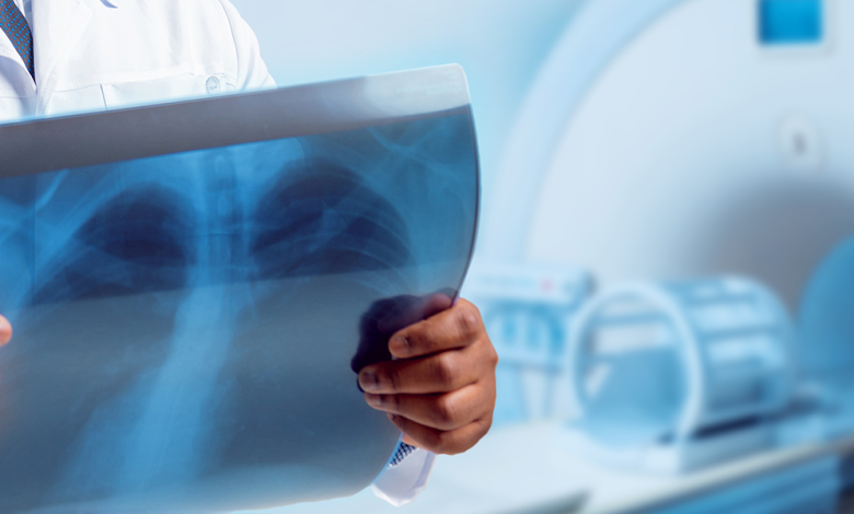
Radiation imaging of the inside of the body was first achieved using X-rays, which have been in use since 1895. As X-rays impact photographic film, they have the additional virtue of darkening the film when they travel through it. For example, bones absorb more X-rays and take longer to show up on film because of the uneven absorption of the rays as they move through tissues.
In addition, while bones absorb many X-rays, soft tissues absorb a much less amount. Consequently, when an X-ray images the inside of the body, bones appear as brighter parts, and soft tissues appear as darker areas on the exposed film.
It’s tough to tell the difference between surrounding soft tissues of almost the same density when X-rays are all that’s being used. However, when an X-ray contrast medium, a liquid or gaseous substance, is injected into the body, the X-rays may be seen clearly.
In some instances, a contrast medium fluid may be injected into naturally occurring body cavities, delivered into the circulatory and lymphatic arteries, swallowed or given via an enema for digestive system studies, or injected around organs to reveal their external form.
Mixed contrast media enable the X-ray imaging of specific soft internal structures. Using X-rays makes it possible to analyze almost any bodily region for physiological irregularities. This includes the passage of blood through the heart in angiocardiography and the gallbladder in cholecystography. Motion picture films may capture the processes inside the body and record certain body sections. X-ray motion picture films may capture the processes inside the body and record them.
One can build another imaging system through X-rays. For example, radiologists may get pictures of deep inside organs and structures through tomography by directing radio waves to a precise plane inside the body. The CT scan services in NJ, also known as computed tomography, is a more advanced technology.
Computed tomography (CT) scan
You can see cross-sectional views of the body with CT scans, also known as CAT scans. An X-ray with cross-sectional pictures provides more detailed views than an X-ray with conventional images.
The patient is placed in the middle of a big doughnut-shaped machine, and while the CT scanner collects pictures, the patient moves through the center. A contrast dye may be injected or swallowed by the patient during specific examinations to assist the doctor see inside the body. At this point, the technician may depart and return when everything is ready to scan. The technician controls the scanner, which carries the patient gently through the control room.
Nuclear medicine
In nuclear medicine, scan radioactive isotopes delivered into the body for any remaining activity. Brain scans involve both isotope scanning and X-ray imaging. Positron emission tomography, a kind of imaging closely associated with isotope scanning, is one such diagnostic imaging method.
Nuclear magnetic resonance (NMR) is another diagnostic imaging technique that uses high-frequency radio waves to take pictures of tiny body slices. In addition, physicians can detect internal organ problems by using high-frequency sound waves known as ultrasounds. Diagnostic imaging uses a wide variety of radiation and various procedures for utilizing them.
Endoscopy and related procedures
Radiologists use flexible optical tools in procedures, including endoscopy, laparoscopy, and colposcopy. They insert these devices into the body through natural or surgically created apertures. In addition, physicians and surgeons may use scope equipment equipped with tiny video cameras to investigate the tissues on a large display. As part of the visual analysis process, they use a variety of scopes to collect a small sample of tissue for histological examination.
Magnetic Resonance Imaging (MRI)
Magnetic resonance imaging (MRI) uses superconducting magnets and radio waves. MRI machines use enormous magnets to generate a magnetic field, which detects disease. MRI uses magnetic fields and radio waves to produce pictures of organs and tissues in an MRI scan. Doctors utilize MRI to examine their patients’ organs, soft tissues, and ligaments. A brain MRI may assist physicians in diagnosing several illnesses, such as vision abnormalities, strokes, tumors, and aneurysms.
Patients with heart disease, heart attacks, or blood vessel inflammation may benefit from MRI imaging. As a result, magnetic resonance imaging identifies cancer in the pancreas and other parts of the digestive system.
Vascular Interventional Radiography
Vascular Interventional Radiology (VIR) enables physicians to treat numerous problems by performing angioplasty and other similar procedures. Interventional radiology uses multiple diagnostic imaging methods and approaches in vascular including computed tomography, ultrasound, and X-ray fluoroscopy.
Interventional radiologists often use tiny skin incisions to deliver small needles to treatment sites during vascular interventional radiology treatments. In addition, physicians may use catheter tubes or wires to traverse a patient’s body during certain operations. For example, it is possible to remove gallbladder stones or kidneys using vascular interventional radiology. This treats diseases of the blood vessels and veins and guides the treatment of benign tumors.
Positron Emission Tomography (PET) Scan
Positron emission tomography (PET) scans are like illness detection in the body since they identify abnormalities in cells. Complete procedure using radioactive tracers. Tracers can identify issues that would otherwise go unnoticed until they deteriorate.
Administered tracers in one of three methods, depending on the procedure: intravenously, by inhalation of gas, or orally via a particular combination. One has to wait for an hour-long since tracers take time to go through the body. Place the patient on a table that slides into an O-shaped machine when it is time. Medical practitioners instruct patients to hold their breath and to remain still by the technician.


